

Published on Nov 30, 2023
Optical Coherence Tomography (OCT) is an imaging technique that is similar in principle to ultrasound, but with superior resolution. It relies on exposing a sample to a burst of light and then measuring the reflective response from different depths and is therefore capable of scanning non-invasively beneath the surface of the sample. In ultrasound imaging, it is relatively easy to measure the time delay of each reflected packet.
However, for light pulses, interferometry must be used to measure the displacement with meaningful accuracy. The amount of light reflected from each point within the scanning window in the sample is plotted graphically as an OCT image.
The goal of this investigation is to use Optical Coherence Tomography to image epileptic lesions on cortical tissue from rats. Such images would be immensely useful for surgical purposes. They would detail how deep the lesion is, allowing for precise removal that neither removes an insufficient amount of damaged tissue nor extracts too much healthy tissue.
Though commerical OCT systems already exist, they typically do not scan very deeply beneath sample surfaces. For the purpose of this study, a system must be constructed that scans up to 2 millimeters into tissue1. Unfortunately, an increase in axial depth necessitates a decrease in transverse (along the surface of the sample) resolution due to focal restrictions of the objective lenses2. However, this loss is acceptable for this investigation, as the main goal is to determine lesion depth and not to achieve perfect image clarity.
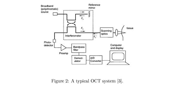
The ability to detect the positional delay of light reflecting from a tissue sample is at the heart of OCT. Low-coherence interferometry provides just that. A low-coherence light source has the potential to produce interference fringes only when integrated with light from the same source that has traveled nearly exactly the same distance3. This means that if light from such a source is split by a beam splitter into two equal parts, both of them reflect off of different objects, and combine to form one beam again, they will produce an interference fringe pattern only if thedistance they traveled while split was exactly the same.
The main application of OCT is in the situations where other techniques are either hazardous or of limited value, in addition to its feasibility for micro-surgical procedures .
In many occasions, for instance in brain, the conventional biopsy method can be extremely dangerous. The competing techniques could also be of limited value due either to their low resolution or in situations with high false negative rates. The detection of many retinal diseases in early stages when still cure methods is useful requires micrometer scale imaging of eye structure which up to now, no other techniques except OCT has been able to accomplish. There are also situations where due to the inherent nature of the disease, there is a great probability of missing the correct diagnosis, though the physician was looking for it; these are referred to as false negative rates.
There are also other types of OCT which are also utilize light to produce imaging of tissue however they tend to include or vary the components of the system to provide more emphasis and extract more information from scans based on something specific from the sample. One of these ideas is the Doppler OCT which looks from for frequency shifts in the interference patterns which would show moving objects in the sample such as blood cells. This is particularly interesting to ophthalmologists since variations in blood flow can be causes of blindness, including diabetic retinapothy and macular degeneration . In addition to Doppler OCT, researchers are also looking into polarization which would measure the polarization of returning light and interference fringes since this might be a way to image damage to tissue such as nerve fibers, skin, and other connective tissues (Rollins 2005).
Polarization OCT might also prove to be helpful in assessing burn severity since the burns result in denaturing of connective tissue proteins and it has been shown to detect differences between healthy and damaged tissues (Rollin 2005). One other form of OCT that is being investigated is the spectroscopic OCT which examines how much light returns from tissues and how the spectrum of that light changes (Rollin 2005). This should enable physicians to characterize the metabolic state or biochemistry of the tissue.
The numerous applications that OCT can be used for is remarkable. OCT has the potential to be used for a great number of medical fields and applications however, cancer and heart disease are two of the most pressing and promising areas of application The imaging from the optical coherence tomography has the potential to improve the current cardiovascular therapies and procedures such as stenting and balloon angioplasty by means of providing vascular images in real time to guide stent placement and balloon inflation. Since OCT has the capability of clearly identifying plaques in the blood stream thus being able to differentiate between stable plaques and unstable plaques, which are probably responsible for up to seventy percent of all heart attacks, it aids those that suffer from cardiac problems. In addition to helping those with cardiac problems, OCT can also be valuable others.
OCT can also potentially improved the current conventional method of biopsies by being more precise in identifying and defining the areas that should be removed or left alone based on the images fro the epithelial layers. This in turn will reduce the number of biopsies and also make diagnosis more accurate and faster. Hopefully as technology becomes increasingly more advanced, it will be possible for medical physicians to perform biopsies only using optical coherence tomography imaging.
In terms of other medical imaging devices, OCT is one the best offered currently.
Optical coherence tomography has becoming a rapidly important biomedical tool for imaging tissues. OCT has critical advantages over other medical imaging systems. Microscopes work well for examining small tissue smalls and cells but not for examining biological tissues inside the body . The Ultrasound, CT, and MRI can peer inside the body however they do not have the resolution or depth to capture cellular detail. Electron microscopy can pick up extremely fine detail however; it is not able to view living samples within the body . The two major advancements which assist both scientists and medical physicians is the improvement in the depth and clarity at which they can view tissues. “Current OCT systems have resolutions at 4-20 um compared to 110 um for high frequency ultrasound”. The advancement of medical imaging systems is illustrated by the technology inherent in OCT, which allows images of living tissue to become clearer and more refined in both structure and detail.
“Using information inherent to the returning photon signals, OCT can perform both spectroscopic and polarization imaging to better evaluate the composition of tissues and lesions” . Another technological advancement which makes OCT that much easier to integrate into potential human subjects is having a small enough device to utilize OCT within the human body. The use of fiberoptics and the ability to combine with catheters allows optical coherence tomography to access the small parts of the body. With other technology to aid the development of OCT, doctors can actually use OCT not only on the body but within the body.
Optical coherence tomography (OCT) has been emerged a novel diagnostic tool for bio medical applications, especially in situations where conventional imaging methods are either hazardous or of little valuable information. The continuing success of OCT depends on the design and fabrication of cost effective as well as portable ultra wide-band light sources in order to pro¬vide better resolution. The development of a firm theoretical basis together with sophisticated digital signal processing algorithms for combating the non-idealities at a post-processing level is apparently a must
| Are you interested in this topic.Then mail to us immediately to get the full report.
email :- contactv2@gmail.com |
