

Published on Apr 02, 2024
Visualizable objects in biology and medicine extend across a vast range of scale, from individual molecules and cells through the varieties of tissue and interstitial interfaces to complete organs, organ systems, and body parts.
The practice of medicine and study of biology have always relied on visualizations to study the relationship of anatomic structure to biologic function and to detect and treat disease and trauma that disturb or threaten normal life processes. Traditionally, these visualizations have been either direct, via surgery or biopsy, or indirect, requiring extensive mental reconstruction. The potential for revolutionary innovation in the practice of medicine and in biologic investigations lies in direct, fully immersive, real-time multi sensory fusion of real and virtual information data streams into online, real-time visualizations available during actual clinical procedures or biological experiments. In the field of scientific visualization, the term "four dimensional visualization" usually refers to the process of rendering a three dimensional field of scalar values.
"4D" is shorthand for "four-dimensional"- the fourth dimension being time. 4D visualization takes three-dimensional images and adds the element of time to the process. The revolutionary capabilities of new three-dimensional (3-D) and four-dimensional (4-D) medical imaging modalities along with computer reconstruction and rendering of multidimensional medical and histologic volume image data, obviate the need for physical dissection or abstract assembly of anatomy and provide powerful new opportunities for medical diagnosis and treatment, as well as for biological investigations.In contrast to 3D imaging diagnostic processes, 4D allows doctor to visualize internal anatomy moving in real-time. So physicians and sonographers can detect or rule out any number of issues, from vascular anomalies and genetic syndromes. Time will reveal the importance of 4d visualization
"4D" is shorthand for "four-dimensional"- the fourth dimension being time. 4D visualization takes three-dimensional images and adds the element of time to the process.
In contrast to 3D imaging diagnostic processes, 4D allows doctor to visualize internal anatomy moving in real-time. For example: Movement patterns of fetuses allows conclusions to be drawn about their development; increase of accuracy in ultrasound guided biopsies thanks to the visualization of needle movements in real time in all 3 planes. So physicians and sonographers can detect or rule out any number of issues, from vascular anomalies and genetic syndromes
Locked within 3-D biomedical images is significant information about the objects and their properties from which the images are derived. Efforts to unlock this information to reveal answers to the mysteries of form and function are couched in the domain of image processing and visualization. A variety of both standard and sophisticated methods have been developed to process (modify) images to selectively enhance the visibility and measurability of desired object features and properties. For example, both realism-preserving and perception-modulating approaches to image display have significantly advanced the practical usefulness of 4-D biomedical imaging.
Many life-threatening diseases and/or quality-of-life afflictions still require physical interventions into the body to reduce or remove disease or to alleviate harmful or painful conditions. But minimally invasive or noninvasive interventions are now within reach that effectively increase physician performance in arresting or curing disease; reduce risk, pain, complications, and reoccurrence for the patient; and decrease healthcare costs. What is yet required is focused reduction of recent and continuing advances in visualization technology to the level of practice, so that they can provide new tools and procedures that physicians ‘‘must have’’ to treat their patients and empower scientists in biomedical studies of structure-to function relationships.
Forming an image is mapping some property of an object onto image space. This space is used to visualize the object and its properties and may be used to characterize quantitatively its structure or function. Imaging science may be defined as the study of these mappings and the development of ways to better understand them, to improve them, and to use them productively. The challenge of imaging science is to provide advanced capabilities for acquisition, processing, visualization, and quantitative analysis of biomedical images to increase substantially the faithful extraction of useful information that they contain.
In the field of scientific visualization, the term "four dimensional visualization" usually refers to the process of rendering a three dimensional field of scalar values. While this paradigm applies to many different data sets, there are also uses for visualizing data that correspond to actual four-dimensional structures. Four dimensional structures have typically been visualized via wire frame methods, but this process alone is usually insufficient for an intuitive understanding. The visualization of four dimensional objects is possible through wire frame methods with extended visualization cues, and through ray tracing methods. Both the methods employ true four-space viewing parameters and geometry.
The ray tracing approach easily solves the hidden surface and shadowing problems of 4D objects, and yields an image in the form of a three-dimensional field of RGB values, which can be rendered with a variety of existing methods. The 4D ray tracer also supports true four-dimensional lighting, reflections and refractions. The display of four-dimensional data is usually accomplished by assigning three dimensions to location in three-space, and the remaining dimension to some scalar property at each three-dimensional location. This assignment is quite apt for a variety of four-dimensional data, such as tissue density in a region of a human body, pressure values in a volume of air, or temperature distribution throughout a mechanical object
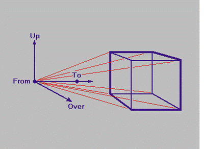
The viewing-angle is defined as for three-dimensional viewing, and is used to size one side of the projection-parallelepiped; the other two sides are sized to fit the dimensions of the projection-parallelepiped. For this work, all three dimensions of the projection parallelepiped are equal, so all three viewing angles are the same.
Raytracing solves several rendering problems in a straight-forward manner, including hidden surfaces, shadows, reflection, and refraction. In addition, raytracing is not restricted to rendering polygonal meshes; it can handle any object that can be interrogated to find the intersection point of a given ray with the surface of the object. This property is especially nice for rendering four-dimensional objects, since many N-dimensional objects can be easily described with implicit equations
For robustly measuring temporal morphological brain changes, a 4D image warping mechanism can be used. Longitudinal stability is achieved by considering all temporal MR images of an individual simultaneously in image warping, rather than by individually warping a 3D template to an individual, or by warping the images of one time-point to those of another time-point. Moreover, image features that are consistently recognized in all time-points guide the warping procedure, whereas spurious features that appear inconsistently at different time-points are eliminated. This deformation strategy significantly improves robustness in detecting anatomical correspondences, thereby producing smooth and accurate estimations of longitudinal changes. The experimental results show the significant improvement of 4D warping method over previous 3D warping method in measuring subtle longitudinal changes of brain structures.
4D-HAMMER, involves the following two steps:
(1) Rigid alignment of 3D images of a given subject acquired at different time points, in order to produce a 4D image. 3D-HAMMER is employed to establish the correspondences between neighboring 3D images, and then align one image (time t) to its previous-time image (t-1) by a rigid transformation calculated from the established
correspondences.
(2) Hierarchical deformation of the 4D atlas to the 4D subject images, via a hierarchical attribute-based matching method. Initially, the deformation of the atlas is influenced primarily by voxels with distinctive attribute vectors, thereby minimizing the chances of poor matches and also reducing computational burden. As the deformation proceeds, voxels with less distinctive attribute vectors gradually gain influence over the deformation
Advanced medical imaging technology allows the acquisition of high resolved 3D images over time i.e.4D images of the beating heart. 4D visualization and computer supported precise measurement of medical indicators (ventricle volume, ejection fraction, wall motion etc.) have the high potential to greatly simplify understanding of the morphology and dynamics of heart cavities, simultaneously reduce the possibility of a false diagnosis. 4D visualization aims at providing all information conveniently in single, stereo, or interactively rotating animated views.
The goal of the 2nd year of the Med-SANARE project is twofold. On one hand a virtual table metaphor will be utilized to set up a visionary high-end cardiac diagnosis demonstrator for educational purpose that makes use of augmented reality (AR) techniques. On the other hand a Cardiac Station will be implemented as functional reduced solution that supports image evaluation making use of standard PC-based technology. The functionality offered will be sufficient to successfully perform the tasks required by the diagnostic procedure. For both systems realistic and detailed modeling and visualization plays a crucial role
W. de Leeuw and R. van Liere. Case Study: Comparing Two Methods for Filtering External Motion in 4D Confocal Microscopy Data. Joint Eurographics .
W.E. Lorensen and H.E. Cline. Marching cubes: a high resolution 3D surface construction algorithm. Computer Graphics (Siggraph’87 Proceedings), 21(4), pp163-169.
M. Levoy. Display of surfaces from volume data. Computer Graphics & Application
| Are you interested in this topic.Then mail to us immediately to get the full report.
email :- contactv2@gmail.com |
